
All living things keep the solution composition ratio constant inside the cells, especially the concentration of ions is critically controlled. When there is irritation from the outside, this solution composition ratio is temporarily disturbed, and it can be felt as stimulation. To recover the solution composition ratio, the cell uses an energy of ATP to control the concentration of ions in the cell. One of the proteins that play this role is V-ATPase.
The Eh V-ATPase used in this study is a V-ATPase present in the cell membrane of Enterococcus hirae, and selectively excretes sodium ions to outside of the cell. The entire structure of Eh V-ATPase was unknown, where the partial structures of Eh V-ATPase was only revealed by X-ray crystallographic structural analysis. This was because the particle image of the solubilized Eh V-ATPase was not able to be visualized by conventional cryo-electron microscopy which was the only way to see the entire structure of the complex.
In this study, we succeeded in visualizing the particle image of Eh V-ATPase solubilized from cell membrane for the first time, using Zernike phase contrast cryo-electron microscopy developed at the National Institute for Physiological Sciences. Then, the entire three-dimensional structure of Eh V-ATPase was reconstructed by single particle analysis by using a number of two-dimensional particle images taken from various angles. V-ATPases generally show machine-like structure in which a rotary motor driven by ATP hydrolysis energy called V1 and another ion-pump rotary motor called Vo are connected by a single rotor. Here, we revealed the unique features of Eh V-ATPase, where the V1 and Vo domains are fixed with two stalks, and the central rotor extending from V1 is connected to the ring of Vo rotor with the off-axis manner. Because of the off-axis connection, the rotation of the ring is expected to be highly eccentric. In addition, the structure artificially fixed the central rotor by binding the antibody revealed the second stable structure among three consecutive structural states of this enzyme, thereby the images of two of three stages of pumping motion were successfully captured.
Based on these results, we could clarify the structural basis of energy transduction mechanism for sodium ion transport of Eh V-ATPase. By further structural research, it is expected that it will be able to provide important information in the treatment of diseases related to V-ATPase and development of therapeutic drugs in the future.
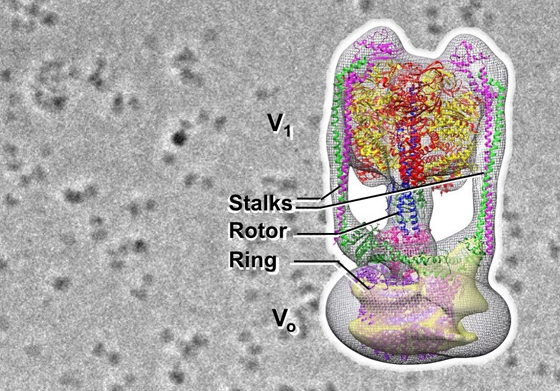
Figure: Image of Eh V-ATPase by Zernike phase contrast cryo-electron microscopy (background) and the 3D structure
Research Group
Ryota Iino (Institute for Molecular Science)
Fabiana Lica Imai, Takeshi Murata (Chiba University)
Hiroshi Ueno (University of Tokyo)
Junichi Takagi (Osaka University)
Jun Tsunoda, Chihong Song, Kazuyoshi Murata (National Institute for Physiological Sciences)
Financial Supports
KAKENHI JP15H04366, JP26102507, JP18H05424, JP16K07282, JP16H06280, JP17H03638, JP18H05425
Advanced Bioimaging Support (ABiS)
JST-ERATO Momose Quantum-beam Phase Imaging Project
Collaborative Study Program of the National Institute for Physiological Sciences
Information of the paper
Title: Off-axis rotor in Enterococcus hirae V-ATPase visualized by Zernike phase plate single-particle cryo-electron microscopy
Authors: Jun Tsunoda, Chihong Song, Fabiana Lica Imai, Junichi Takagi, Hiroshi Ueno, Takeshi Murata, Ryota Iino, Kazuyoshi Murata
Journal: Scientific Reports
Issue: 8:15632
Date: 2018.10.23 publish & online
URL (abstract): https://www.nature.com/articles/s41598-018-33977-9
doi: 10.1038/s41598-018-33977-9
2910
