English version 26 may, 2020 on EurekAlert!
A new imaging electron analyzer that visualizes the electronic structure of micrometer-scale functional materials in both real space and momentum space is now in operation at the third-generation synchrotron facility UVSOR-III
The importance of characterizing electronic properties of μm- and nm-scale world cannot be overemphasized. A new experimental station for 3D momentum-resolved photoelectron spectroscopy is constructed at UVSOR synchrotron facility. Microscopy with a spatial resolution of 50 nm and photoelectron spectroscopy (field of view: 2 μm) with energy / momentum resolutions of 20 meV / 0.01 Å-1 at 9 K are successfully demonstrated. A method to directly observe the electronic structure of μm-sized materials are realized.
A new momentum microscopy experimental station for photoelectron spectroscopy resolved in 3D momentum space with a microscopic field of view has been built at BL6U of UVSOR*, Institute for Molecular Science. The momentum microscope opens the door to direct observation of the Fermi surface and band structure of μm-sized targets such as surface atomic sites, thin films and interfaces, molecular adsorbates, and polycrystals, which was difficult with conventional electron energy analyzers.
Understanding based on the atomic level characterization methods is becoming increasingly important in elucidating new quantum phenomena and developing functional materials and devices. In photoelectron spectroscopy (PES), the photoelectrons emitted from the sample surface irradiated with X-rays are measured to clarify the composition and electronic structure of the sample. So far, high-resolution electron analyzers have been developed to measure the valence band dispersion of crystalline samples, but they average information over a wide field of view. However, the most interesting materials and useful devices are often micrometer-scale polycrystalline or highly integrated structures. Therefore, a high-specification electron energy and momentum analysis apparatus that also has microscope functions was desired. The momentum microscope, a combination of projection-type electron analyzer and photoelectron microscope, simultaneously realizes a microscope function for magnifying and observing minute parts of complicated-structured samples with element selectivity and a spectroscopy function for visualizing electron behavior (momentum) that determines the electronic properties of a sample. Now we have introduced the latest momentum microscope at the undulator-based soft X-ray beamline of UVSOR providing the optimal energy light for valence bands and core levels of various elements. A spatial resolution of 50 nm for microscopy measurement were achieved. PES measurement with a momentum resolution of 0.01 Å-1 and an energy resolution of 20 meV were successfully demonstrated. The smallest field of view for PES is 2 μm in diameter. In addition to the features of momentum microscope, the sample temperature can be freely changed from 9 K (-264 ºC) to 400 K (127 ºC), which enables in-situ observation of phase transition in the materials. One can investigate how the composition, structure, and electronic state of materials are related to electronic properties and functions.
The momentum-resolved micro-photoelectron spectroscopy realized by this device is an important method for developing nanomaterial science and quantum device engineering through electronic structure analysis. It enables electronic property and function analysis from a new point of view, such as direct observation of structure and electronic state changes where superconductivity and catalytic activity are occurring. In addition to contributing to basic science and applied research, we aim to make this new pioneering analyzer and analysis method a precursor to measurement technology innovation. This work was published online in Japanese Journal of Applied Physics on May 21.
* UVSOR Synchrotron Facility:
UVSOR is a synchrotron radiation facility at Institute for Molecular Science, Japan with the world’s highest performance in the extreme ultraviolet energy range and is widely used by domestic and overseas researchers. The extreme ultraviolet energy range is suitable for observing behavior of electrons which are responsible for properties of molecules and solids. Synchrotron radiation emitted from an electron storage ring with a circumference of about 50 meter is introduced into more than a dozen experimental stations in which a wide variety of researches on bioscience, environmental and energy sciences as well as physical and chemical sciences are conducted. Although it is the second oldest synchrotron radiation facility in Japan since the first light was observed in 1983, it successfully keeps state-of-the-art performance with undergoing intensive upgrades twice.
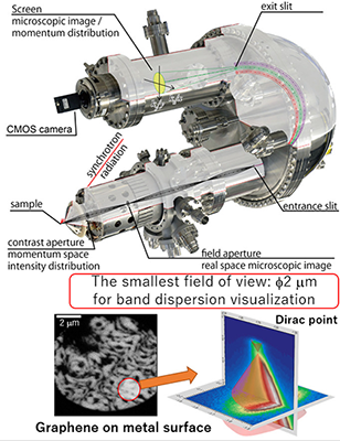
Figure 1: The upper part is a schematic diagram of a photoelectron momentum microscope. The spatial and momentum distribution of photoelectrons emitted from the sample surface by irradiation with synchrotron radiation are analyzed and projected on the screen. An example of a real-space image of graphene grown on a metal surface is shown. The field of view can be changed within the range of 200 to 2 μm in diameter. A 50 nm spatial resolution is achieved. One can select the region of interest and visualize the electronic structure by switching the system from microscope to spectroscopic mode.
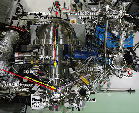
Figure 2: Drone photo of a photoelectron momentum microscope installed in the experimental station at the soft X-ray beamline BL6U of the UVSOR-III synchrotron facility is shown. The system is equipped with various excitation sources, sample preparation equipment, and storage.
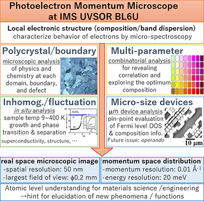
Figure 3: The specifications of UVSOR-III photoelectron momentum microscope are summarized. The momentum microscope opens the door to direct observation of the Fermi surface and band structure of μm-sized targets such as surface atomic sites, thin films and interfaces, molecular adsorbates, and polycrystals. The momentum-resolved micro-photoelectron spectroscopy realized by this system is an important method for developing nanomaterial science and quantum device engineering through electronic structure analysis. One can investigate how the composition, structure, and electronic state of materials are related to electronic properties and functions. It enables electronic property and function analysis from a new point of view, such as direct observation of structure and electronic state changes where superconductivity and catalytic activity are occurring.
Information of the paper:
Authors: Fumihiko Matsui, Seiji Makita, Hiroyuki Matsuda, Takayuki Yano,
Eiken Nakamura, Kiyohisa Tanaka, Shigemasa Suga, Satoshi Kera
Journal Name: Japanese Journal of Applied Physics
Journal Title: “Photoelectron Momentum Microscope at BL6U of UVSOR-III Synchrotron”
DOI: https://doi.org/10.35848/1347-4065/ab9184
Financial Supports:
Grant-in-Aid for Scientific Research (19KK0137, 18H03904, 17H02911)
Contact Person:
Fumihiko Matsui
TEL: +81-564-7201
E-mail: matui_at_ims.ac.jp *Please replace the "_at_" with @
1594
1705
3907
