Executive summary
● IMS research team observed a single protein through infrared light confined to the nanometer scale.
● Using the infrared near-field microscopy, the team demonstrated infrared vibrational spectroscopy of a single protein.
● The team also theoretically elucidated the interaction between infrared light and proteins in nanocavity.
Overview
Infrared vibrational spectra, often referred to as “molecular fingerprints”, are widely used for the structural and chemical analysis of various materials. The rapid development of nanotechnology in recent years has led to increasing demand for ultra-high sensitivity and super-resolution infrared imaging. However, conventional infrared spectroscopy is limited in terms of its sensitivity and spatial resolution. For example, even the best infrared microscope requires over a million proteins within tens of micrometer volume for obtaining an infrared vibrational spectrum, rendering it impossible to measure just a single protein.
An interdisciplinary research team, led by Jun Nishida (Assistant Prof.) and Takashi Kumagai (Associate Prof.) at Institute for Molecular Science, has successfully observed infrared vibrational spectra of single proteins using infrared near-field optical microscopy (Note 1). This method utilizes light confined at the nanometer scale, allowing for the detailed analysis of extremely small samples, which was challenging with conventional infrared spectroscopy.
In their study, the research team isolated a single protein, a sub-unit comprising a protein complex called F1-ATPase (Note 2), on a gold substrate and performed near-field infrared spectroscopy measurements in an ambient environment. They successfully acquired the infrared vibrational spectrum of a single protein, representing a major advance that may lead to characterizing local structural organizations of individual proteins. Such information is particularly important for understanding the sophisticated functions of protein complexes and membrane proteins, offering deeper insights into their mechanisms and interactions. Furthermore, they have developed a new theoretical framework describing the nanoscale interactions between the infrared near field and protein. Based on the theory, the team was able to quantitatively reproduce the experimental vibrational spectra that they observed. These results will be invaluable for the chemical analysis of biomolecules as well as various nanomaterials, paving the way for a range of applications of nanoscale infrared spectroscopy.
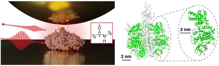
Figure 1: (Left) Scheme of near-field infrared spectroscopy measuring a single protein. (Right) The structure of the protein complex F1-ATPase and the subunit measured in this study.
Details
Infrared near-field optical microscopy (known as nano-FTIR, Note 3) has emerged and rapidly established itself in recent years as an ultra-sensitive and super-resolution imaging technique in the infrared regime. Nano-FTIR enables the characterization of material composition and structure at the nanometer scale—a capability that is significant in both fundamental scientific research and various industrial applications. Nano-FTIR has found widespread applications in different branches of science, and its technological progress would impact a variety of research fields, from biological science to material engineering, due to its capability to provide unique insights into material properties at the nanoscale.
The team in Institute for Molecular Science has developed a highly sensitive nano-FTIR system and has undertaken the challenge of measuring the infrared vibrational spectra of a single protein consisting only of 500 amino acid residues—a feat previously unachieved. Figure 1 shows a schematic diagram of the nano-FTIR system developed for this study. As a mid-infrared light source, the team employs a pulsed laser source with high stability and optimal bandwidth for vibrational spectroscopy applications. When the apex of a metallic tip in an atomic force microscope (AFM) is illuminated with the infrared pulse, a highly localized field called near field is formed at the nanoscale. The interaction between this near field and a protein deposited on a gold surface results in infrared scattering, which is detected by interfering with another pulse as in Fourier transform infrared spectrometer (FTIR, Note 4) to yield a nanoscale infrared spectrum.
The scattering amplitude sensitively depends on the distance between the tip and the sample. The team took advantage of this unique property by modulating the tip‒sample distance at a frequency ωAFM and detecting the signal component that is modulated at the higher harmonics of the frequency (nωAFM) with a lock-in amplifier (Note 5). This not only allowed the researchers to selectively detect the near-field scattering, but also to have access to highly localized near-field interactions confined to a single-nanometer scale by analyzing the higher harmonics (n) component.
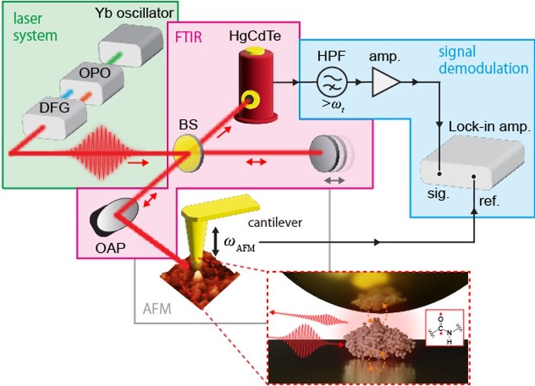
Figure 2: Schematic diagram of the nano-FTIR system developed for this study. Yb oscillator: Ytterbium femtosecond laser, OPO: Optical Parametric Oscillator, DFG: Difference Frequency Generator, BS: Beam Splitter, OAP: Off-Axis Parabolic Mirror, HgCdTe: Infrared Detector, HPF: High Pass Filter, amp.: Preamplifier, Lock-in amp.: Lock-in Amplifier, sig.: Detected Signal, ref.: Reference Signal, ωAFM: Resonant Frequency of the Cantilever.
Figure 3(a) and (b) show the AFM image and infrared spectrum of a single protein, respectively. In the AFM image a profile reflecting a topography of a single protein is observed, and in the infrared spectrum a vibrational resonance originating from amide groups in the protein is detected at 1665 cm-1. The amide vibrations are known to reflect secondary structures of proteins and has been extensively studied based on conventional FTIR spectroscopy and more advanced nonlinear infrared spectroscopy methods. In this work, the team demonstrated that the equivalent information can be acquired at the single protein level.
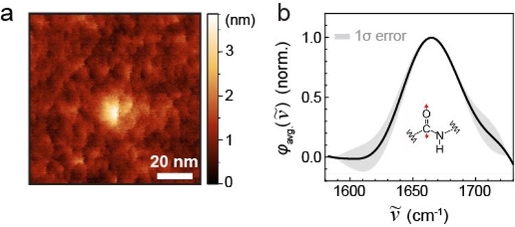
Figure 3: AFM image (a) and infrared spectrum (b) of a single protein.
The research team also demonstrated that the localization of the near-field scattering signal is significantly enhanced by analyzing the higher-order demodulation harmonics using the lock-in amplifier. Figure 4(a) shows a plot of the infrared near-field scattering amplitude against the distance between the tip and the sample in the nano-FTIR. The plot clearly shows that as the order of demodulation harmonics increases, the decay length of the infrared near-field scattering amplitude becomes shorter, indicating a more localized interaction. Additionally, as shown in Figure 4(b), the vibrational resonance in the infrared spectrum becomes stronger at the higher orders. These results demonstrate that higher-order demodulation probes more spatially localized near-field interactions. Figure 4(c) shows a graph plotting the decay length of the infrared near-field scattering amplitude against the order of demodulation, revealing that for n ≥ 6, the infrared near-field signal is localized to less than 5 nm.
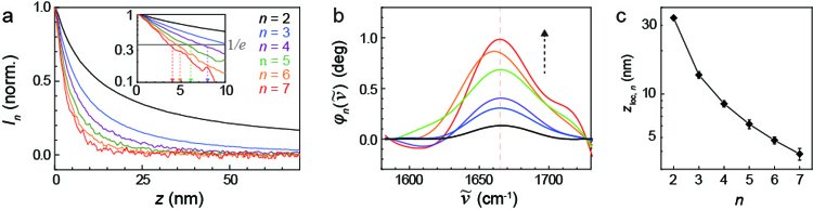
Figure 4: (a) A graph plotting the infrared near-field scattering intensity (vertical axis) against the distance between the tip and the sample (horizontal axis). The graph shows demodulation orders from 2nd to 7th. (b) Infrared spectra measured at demodulation orders from 2nd to 7th. (c) A graph plotting the dependency of the decay length (vertical axis), obtained from the analysis of the graph in (a), against the demodulation order (horizontal axis).
The research team also performed theoretical analysis to rationalize the experimental results, focusing on the interaction between the infrared near field and single protein. The infrared near-field spectrum is obtained by calculating the polarization induced when an electrostatic field is applied to the system. Figure 5(a) depicts the different analytical models that were used for simulations. Figure 5(b) compares the theoretical infrared vibrational to the experimental results. The theory successfully reproduces the enhanced vibrational resonance at the higher demodulation harmonics. The further detailed comparison reveals that the model which closely replicates the actual situation (referred to as EMPD in the study) showed the best agreement with the experimental data.
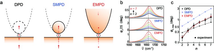
Figure 5: (a) Analytical models used to reproduce the experimental results. DPD: Double Point Dipole Model, SMPD: Spherical Mirror and Point Dipole Model, EMPD: Elliptical Mirror and Point Dipole Model. (b) Calculated infrared spectrum of a single protein. (c) A graph plotting the peak values of the infrared spectrum (vertical axis) against the demodulation order (horizontal axis). Black diamonds: experimental data, black circles: DPD model, red circles: SMPD model, blue circles: EMPD model.
Significance of the Research Findings
Infrared spectroscopy is a versatile method used in physical, chemical, biological, and medical sciences for investigating structures, properties, and functions of materials. With the advancement of nanotechnology, nanoscale spectroscopy techniques have become increasingly important. This research represents a significant step towards the next generation of ultra-sensitive and high-resolution imaging using mid-infrared infrared light, which holds promise for applications in a variety of nanomaterials.
Terms
(Note 1) Near-field optical microscopy: A technique that uses near-field light (nanoscale light) generated at the apex of a sharp metallic tip. Unlike conventional optical microscopes, which are limited in resolution due to the diffraction limit, near-field optical microscopy allows observation of materials at much higher spatial resolutions.
(Note 2) F1-ATPase: A protein complex composed of multiple subunits, which is important in cellular energy metabolism. By rotating like a motor, it synthesizes adenosine triphosphate (ATP), functioning as a cellular energy currency.
(Note 3) Nano-FTIR spectroscopy: Conventional infrared spectroscopy cannot observe materials at scales smaller than micrometers due to the diffraction limit. Infrared near-field optical microscopy can achieve nanometer-scale resolution by observing samples with the near field generated by illumination on a sharp metal probe. When combined with atomic force microscopy (AFM), it can simultaneously capture the structure of the sample and optical information in the infrared region at the nanoscale.
(Note 4) Fourier transform infrared spectrophotometer (FTIR): An instrument that measures the amount of infrared light transmitted or reflected by a sample.
(Note 5) Lock-in amplifier: An electronic device used to detect and amplify very weak signals. It is a sensitive device that can selectively detect and amplify weak components of a specific frequency in an analog signal. Widely used in weak signal detection, it enables accurate measurements even in noisy environments.
Presenter and Researcher Information:
National Institutes of Natural Sciences, Institute for Molecular Science
Jun Nishida (Assistant Professor)
Akihiro Otomo (Assistant Professor)
Takanori Koitaya (Assistant Professor at the time of the research, currently Associate Professor at Kyoto University Graduate School of Science, Department of Chemistry)
Taketoshi Minato (Senior Researcher)
Ryota Iino (Professor)
Takashi Kumagai (Associate Professor)
Max Planck Society, Fritz Haber Institute
Akitoshi Shiotari (Group Leader)
Publication Information:
(Journal) Nano Letters
(Title) Sub-tip-radius near-field interactions in nano-FTIR vibrational spectroscopy on single proteins
(Authors) Jun Nishida*, Akihiro Otomo, Takanori Koitaya, Akitoshi Shiotari, Taketoshi Minato1, Ryota Iino, and Takashi Kumagai*
(DOI) doi.org/10.1021/acs.nanolett.3c03479
Research Funding:
JST FOREST Program (JPMJFR201J, Japan)
Grants-in-Aid for Scientific Research (JSPS KAKENHI: 19J24684)
Advanced Photon Science Project (01213008 and 01212204)
Emerging Research Support Project (JPMJFR201J)
Contact Information:
(For inquiries about the research, please contact the presenters)
National Institutes of Natural Sciences, Institute for Molecular Science
Jun Nishida
Tel: 0564-55-7412
E-mail: nishida_at_ims.ac.jp
National Institutes of Natural Sciences, Institute for Molecular Science
Takashi Kumagai
Tel: 0564-55-7410
E-mail: kuma_at_ims.ac.jp
(Please replace the "_at_" with @)
4693
