To investigate metal‒ligand delocalization of metal porphyrin complexes in aqueous solutions, this study probes the electronic structures of both the metal and ligand sides using soft X-ray absorption spectroscopy (XAS) at the metal L2,3-edges and N K-edges, respectively. In the N K-edge XAS spectra of the ligands, the C=N π* peaks of cobalt protoporphyrin IX (CoPPIX) show higher energy shifts than those of iron protoporphyrin IX (FePPIX), owing to the different electronic configurations and spin multiplicities of metal porphyrin complexes. These spectra are useful for discussing the central-metal dependence of metal‒ligand delocalization. Meanwhile, the N K-edge XAS of CoPPIX and the inner-shell calculations of different hydration models reveal that CoPPIX maintains its five-coordination geometry in aqueous solution.
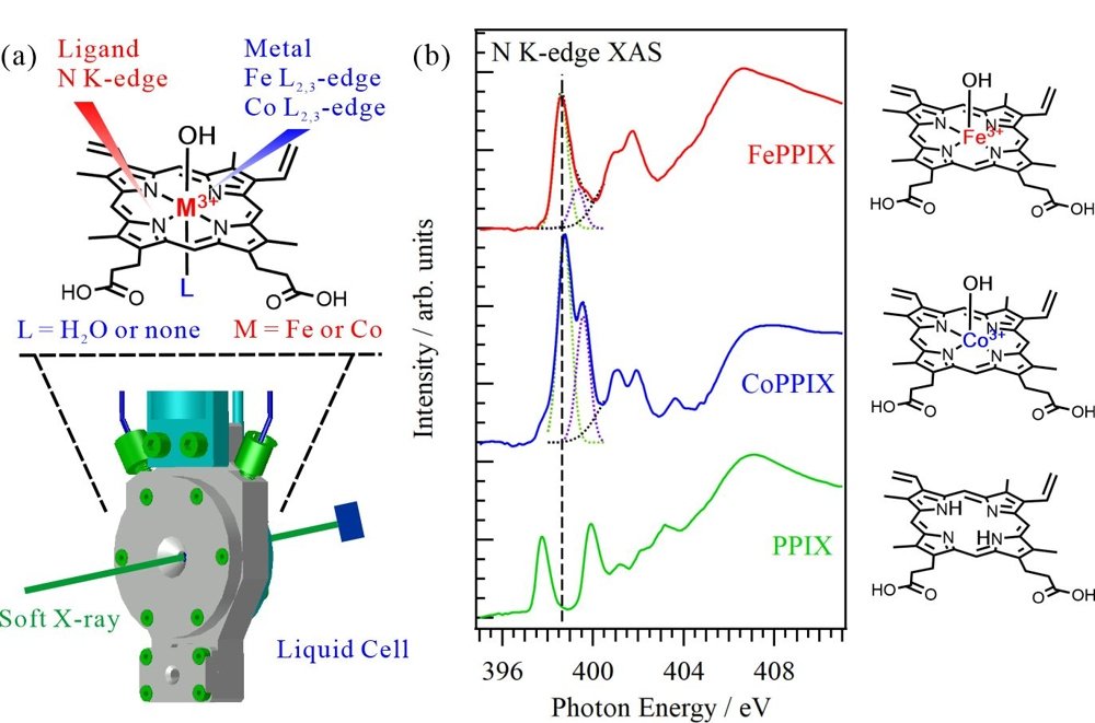
Porphyrin is known as a “pigment of life” because porphyrin and its metal complexes are involved in various biochemical processes such as photosynthetic reactions. The functions of metal porphyrin complexes strongly depend on their molecular structures, which have been extensively investigated in crystal phase through single-crystal X-ray structural analyses. However, many metal porphyrin complexes function not in crystal phases but in solutions or protein scaffolds, where their structures can be dynamically changed. Accordingly, analytical methods enabling direct comparison of the electronic structures and coordination structures of metal porphyrin complexes in solutions or protein scaffolds are required. In this study, the electronic and coordination structures of metal porphyrin complexes in aqueous solutions were investigated using soft X-ray absorption spectroscopy (XAS) of the central metal ions at the metal L2,3-edges and ligands at the N K-edges, respectively, using a liquid-cell apparatus (see Figure 1(a)).

Figure 1: (a) Schematic of the liquid cell for XAS analysis of metal porphyrin complexes in aqueous solutions. XAS spectra of the central metals were obtained at the Fe or Co L2,3-edge and those of ligands were obtained at the N K-edge. The hydration of the metal porphyrin complex was also investigated using N K-edge XAS. (b) N K-edge XAS spectra of FePPIX, CoPPIX, and PPIX in aqueous solutions. The C=N π* peaks of the ligands reflect metal‒ligand delocalization of the metal porphyrin complexes. (Credit:Masanari Nagasaka, Restriction:CC BY)
The XAS experiments were performed at the soft X-ray beamline BL3U of the UVSOR-III Synchrotron. Figure 1(b) shows the N K-edge XAS spectra of 50 mM iron protoporphyrin IX (FePPIX), cobalt protoporphyrin IX (CoPPIX), and protoporphyrin IX (PPIX) in 0.5 M NaOH aqueous solutions. The PPIX spectrum presents two C=N π* peaks contributed by two different types of C=N groups, one connected to H atoms, the other not connected to H atoms. In contrast, the C=N groups of the ligands in FePPIX and CoPPIX are equivalent because four N atoms connect to the metal ion in the same manner. The XAS spectra of FePPIX and CoPPIX show two C=N π* peaks derived from delocalization of the metal 3d orbitals to the N 2p orbitals. The CoPPIX spectrum exhibits a higher first peak and a larger energy difference between its multiple peaks than the FePPIX spectrum. The energy differences of the C=N π* peaks between FePPIX and CoPPIX reflect the different electronic configurations and spin multiplicities of the metal porphyrin complexes. Meanwhile, the N K-edge XAS measurements can effectively observe the central-metal dependence of metal‒ligand delocalization in metal porphyrin complexes. Here, the N K-edge XAS spectrum was obtained for hydrated CoPPIX in aqueous solution. The results, along with inner-shell calculations of different hydration models, revealed that CoPPIX does not form a hydration structure and maintains a five-coordination geometry in aqueous solution.
In summary, N K-edge XAS of ligands can directly compare the electronic structures and coordination structures of metal porphyrin complexes in solutions or protein scaffolds. The present analysis method can explore various chemical and biochemical processes such as photosynthetic reactions, biological redox reactions, and artificial photosynthetic reactions, which involve most metal complexes in solution or protein scaffolds.
Information of the paper:
Authors: Masanari Nagasaka, Shota Tsuru and Yasuyuki Yamada
Journal Name: Physical Chemistry Chemical Physics
Journal Title: “Metal–ligand delocalization of iron and cobalt porphyrin complexes in aqueous solutions probed by soft X-ray absorption spectroscopy”
DOI: https://doi.org/10.1039/D4CP02140A
Financial Supports:
JSPS, KAKENHI, Grant-in-Aid for Scientific Research (B): JP19H02680, JP22H02156
Joint Research by Institute for Molecular Science (IMS): 19-518, 20-719
Alexander von Humboldt Foundation: JPN1201668
Contact Person:
Masanari Nagasaka
Institute for Molecular Science, NINS
Graduate Institute for Advanced Studies, SOKENDAI
TEL/FAX: +81-564-55-7394 / +81-564-55-7493
E-mail: nagasaka_at_ims.ac.jp (Please replace the "_at_" with @)
1058

