| 概 要 |
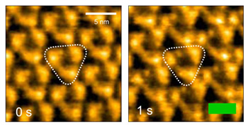
High-speed atomic force microscopy (HS-AFM) is now capable of imaging conformational changes of single proteins in action with sub-second temporal resolution and sub-nanometer resolution. For instance, HS-AFM movie clearly shows the light stimulated conformational changes of bacteriorhodopsin, which functions as the light-driven proton pump through the membrane (Figure) [1, 2]. However, application of HS-AFM in cellular morphology has not yet been demonstrated. In this seminar, I will talk my recent research to apply HS-AFM to living cells, especially neurons. To apply HS-AFM to living neurons, we optimized the original HS-AFM as follows: first, we have developed a long AFM tip (2~3 μm) to avoid collisions between the cantilever and the sample during the imaging. Second, we combined HS-AFM with fluorescence microscopy to locate the AFM tip on the region of interest within neurons. After all these optimizations, we suceeded in imaging HeLa cells and neurons with nanometer resolution with the speed of 1 frames per 5-30 s depending on the scanning region.
[1] M. Shibata et al., Nature Nanotech. 5, 208–212 (2010).
[2] M. Shibata et al., Angew. Chem. Int. Ed. 50, 4410–4413 (2011).
|